
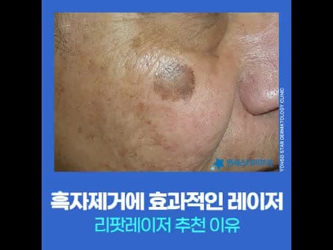
Why Lentigo Pigment Should Be Addressed Early, and RePott Laser
Have you ever experienced small brown spots appearing one by one on your face, only to find their number increasing over time?
First published 2026-06-25 / Last reviewed 2026-07-23
Initially, you might dismiss them, but as they darken and grow in size, becoming difficult to conceal with makeup, this is characteristic of lentigo. Lentigo is not a pigment disorder that disappears on its own, so if you discover it, it is advantageous to address it early when the color is light.
Why Does Lentigo Occur?
Lentigo is primarily caused by chronic cumulative exposure to ultraviolet (UV) light, leading to an excessive proliferation of melanin pigment in the basal cell layer of the skin. It appears as flat, round brown spots, mainly on areas frequently exposed to sunlight, such as the face and back of the hands. It can gradually start appearing as early as the 30s, and tends to increase in number and size with aging and cumulative UV exposure.
Lentigo pigment accumulates in the epidermal layer, making it relatively responsive to laser treatment. However, the older the lesion, the deeper the pigment's roots tend to be, increasing the difficulty of treatment. Therefore, addressing it quickly in the early, lighter-colored stage is easier. Melasma, blemishes, and post-inflammatory hyperpigmentation, which can be confused with lentigo, require different causes and approaches, so differentiation is necessary.
Types and Characteristics of Lentigo
Even though they may look like similar brown spots, lentigo is categorized by its form.
- Simple Lentigo: Small brown spots with clear borders, the most common type, often referred to as blemishes.
- Solar Lentigo: Occurs due to cumulative UV exposure, larger than simple lentigo with distinct borders and deeper roots, posing a risk of recurrence.
- Agminated Lentigo: A form easily mistaken for melasma, appearing as multiple scattered spots.
Sizes are generally flat, round spots of 1-3cm, but there can be individual differences. Lentigo tends to darken in color, increase in size, and spread to surrounding areas over time.
Lentigo Types at a Glance
| Type | Characteristics | Points to Note |
|---|---|---|
| Simple Lentigo | Small brown spots with clear borders | Most common type, often called blemishes |
| Solar Lentigo | Large with distinct borders | Deep roots, potential for recurrence |
| Agminated Lentigo | Multiple scattered spots | Easily mistaken for melasma |
Initially, it's easy to think that light-colored spots will disappear easily, but older lesions tend to have deeper pigment roots, making treatment more challenging. For lentigo, nevus, and spots, it is important to approach treatment after an accurate diagnosis.
Lentigo Laser Treatment: The Principle of RePott Laser
Among the lasers used for lentigo removal, RePott Laser has recently gained attention. RePott Laser is a pigment-specific laser that selectively reacts to melanin pigment, converting absorbed energy into heat to destroy the pigment. It is specialized for epidermal pigment lesions like lentigo and seborrheic keratosis. The actual treatment configuration can be found in the RePott Laser Treatment Guide.
RePott Laser incorporates VSLS vascular protection technology and AutoDerm precision recognition technology, which selectively delivers energy only to pigment lesions while protecting the dermal layer and capillaries.
VSLS technology helps reduce capillary damage and the risk of post-inflammatory hyperpigmentation, which were concerns with existing pigment lasers. Additionally, AutoDerm technology automatically recognizes pigment density and adjusts energy, allowing for an approach tailored to the depth and density of the lesion.
Why Diagnosis Comes First
Various pigment disorders can appear similar to lentigo. Often, melasma, seborrheic keratosis, and freckles are mixed, making accurate differentiation difficult with the naked eye alone. Incorrectly applying high-energy lasers or receiving indiscriminate treatments without a specialist's diagnosis can lead to side effects such as recurrence, worsening hyperpigmentation, or damage to healthy cells.
In clinical practice, it is more common to see lentigo mixed with other pigments rather than appearing alone. Even if they appear to be the same pigment disorder, treatment methods and laser selection can vary depending on the type. Therefore, it is crucial to first identify the nature of the lesion with precise diagnostic equipment before starting treatment. If you have blemishes, seborrheic keratosis, or freckles along with lentigo, Epidermal Pigment Removal can also be considered.
In Summary
If left untreated, lentigo darkens and its roots deepen, making treatment more difficult. Approaching it in the early, lighter-colored stage is a more manageable direction. As lentigo is often mixed with similar pigment disorders, we recommend starting treatment after accurately identifying the lesion with a precise diagnosis. Consult via chat now
Sang-Joo Lee, Chief Director · Dermatology Specialist
As the Chief Director of Yonsei Star Dermatology Sinchon, he has long treated pigment disorders. In 2026, he serves as the President of the Korean Dermatological Association and is also an adjunct professor at the Department of Dermatology, Yonsei University College of Medicine. His profile can be found in About Chief Director Sang-Joo Lee.
First published 2026-06-25 / Last reviewed 2026-07-23
This article provides general medical information, and treatment methods and results may vary depending on individual skin condition and pigment type. For an accurate diagnosis, please consult a dermatology specialist.