
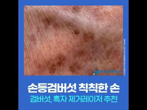
Many people visit the clinic because brown spots on their hands make them look older. If the color has deepened because you haven't paid as much attention to your hands as your face, you can first consider the possibility of seborrheic keratoses.
Why Seborrheic Keratoses Often Appear on the Back of the Hands
Seborrheic keratoses are pigmentary disorders that occur when melanin pigment is excessively deposited and epidermal cells proliferate excessively in skin that has been exposed to accumulated UV radiation for a long time. The back of the hands, along with the face, is the area most exposed to UV light, so seborrheic keratoses often appear there.
As people age, their ability to recover from pigmentation decreases, so the number of lesions often increases significantly after middle age. However, if you have been negligent in UV protection, they can appear at an earlier age.
They Don't Disappear on Their Own, and Increase if Left Untreated
The most significant characteristic of seborrheic keratoses is that their number increases and their color deepens over time. Since they do not naturally lighten or disappear, if brown spots begin to appear on the back of your hands, it is advantageous to check for pigmented lesions such as seborrheic keratoses and lentigo at an early stage.
Most of the time, seborrheic keratoses on the back of the hands increase in number while people postpone treatment, thinking, "they'll disappear if I just leave them alone." In the clinic, the treatment plan for someone who came when there was only one lesion is distinctly different from someone who came after several had spread.
Even if They Look Similar, Pigmentary Disorders Are Different Types
In addition to seborrheic keratoses, there are various types of pigmentary troubles that appear on the back of the hands, such as lentigo, blemishes, melasma, and freckles. Even if they look similar, the skin layer where the pigment is located and the appropriate equipment differ, so accurately identifying the type of pigment is the starting point of treatment.
| Pigment Type | Main Layer | Appearance Characteristics |
|---|---|---|
| Seborrheic Keratosis | Epidermis | Often slightly raised above the surface and thick |
| Lentigo | Epidermis | Flat, clearly defined brown spots |
| Melasma | Epidermis·Dermis | Spreads widely with blurred borders |
| Freckles | Epidermis | Small, light brown spots scattered |
Seborrheic keratoses often appear slightly raised above the surface, and their color can range widely from light brown to almost black. However, not all seborrheic keratoses look the same, so it is safer to confirm an accurate differential diagnosis through a dermatologist's examination.
Why You Shouldn't Pick Them Off with Your Hands
Since seborrheic keratoses are raised, some people try to remove them with their hands or unsterilized tools. This can worsen skin damage and lead to secondary infections, potentially prolonging the treatment period.
At a dermatology clinic, after checking the nature of the lesion and the skin condition, the appropriate laser is selected and performed hygienically, allowing for a faster and more accurate approach than self-treatment.
RePott Laser That Reads and Adjusts Pigment Density
RePott Laser is an equipment whose utilization has recently increased in the treatment of seborrheic keratoses. It is a pigment-specific laser that selectively reacts to melanin pigment, converting absorbed energy into heat to act on pigment cells. It is approved by the Ministry of Food and Drug Safety and is used for epidermal pigmentary lesions such as seborrheic keratoses and lentigo.
Seborrheic keratoses often have varying pigment densities across different areas. The AutoDerm technology applied to RePott Laser recognizes the pigment density of the lesion in real-time and automatically adjusts the energy, making it possible to uniformly approach lesions with varying densities. In addition, a technology that cools and protects blood vessels is added, delivering energy selectively only to the pigmented lesion while protecting the dermis and capillaries during the procedure.
This method helps reduce the burden of post-treatment erythema or pigmentation, which was a concern with existing pigment lasers. If blemishes and freckles are present along with seborrheic keratoses, Epidermal Pigment Removal may be combined, using different equipment for each type of lesion.
Pre-Treatment Differentiation and Surface Preparation
Since seborrheic keratoses can be raised, a process of preparing the lesion surface may be necessary before laser treatment. This is because the surface condition must be refined first for the laser energy to be accurately delivered into the lesion.
Also, treatment should begin after accurately differentiating whether the hand pigmentation is seborrheic keratoses, lentigo, or other pigmentation. Even if they look like the same pigment, choosing the wrong laser can worsen pigmentation or increase the risk of recurrence, so confirmation using precise diagnostic devices like dermoscopy is prioritized.
Post-Treatment Management to Delay Recurrence
Seborrheic keratoses can recur or new lesions can appear if UV exposure continues after treatment. To maintain the results for a long time, it is recommended to consistently follow the following management:
- Apply sunscreen 30 minutes before going out, thoroughly covering the back of the hands.
- Reapply every 2-3 hours during prolonged outdoor activities.
- Do not forcibly remove scabs immediately after the procedure; wait for natural shedding.
- During the recovery period, temporarily stop irritating home care products with whitening or exfoliating ingredients.
- Moisturize sufficiently to help restore the skin barrier.
Frequently Asked Questions
Do seborrheic keratoses disappear in one go? It depends on the thickness of the lesion and pigment density. Shallow lesions may be cleared in relatively early sessions, but if they are thick or dark, additional procedures are planned while observing the progress.
Is the treatment for the back of the hands the same as for the face? The principle of approaching pigmented lesions is similar, but the skin on the back of the hands is thin and recovers slowly, so UV management is particularly important.
If you have any questions, please feel free to inquire via chat now, and we will guide you on the appropriate direction for your condition.
Chief Director Lee Sang-joo · Dermatologist Chief Director of Yonsei Star Dermatology Sinchon and President of the Korean Academy of Dermatology in 2026, he also serves as an adjunct professor in the Department of Dermatology at Yonsei University College of Medicine. He has long specialized in the fields of pigmentation and scars. → Chief Director Lee Sang-joo's Profile and Consultation Guide
First published July 16, 2026 · Last reviewed July 23, 2026
This article provides general medical information, and treatment methods and results may vary depending on individual skin conditions. Please confirm the presence of seborrheic keratoses and your treatment plan through a dermatologist's examination.