
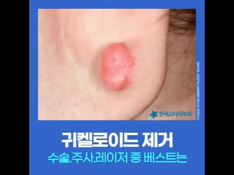
If, after getting an earring or piercing, a lump-like growth appears on your earlobe or helix, doesn't shrink over time, and is accompanied by pain or itching, you might suspect an ear keloid.
First published 2026-06-19 / Last reviewed 2026-07-23
Why Do Keloids Form?
Keloids are scar disorders where collagen excessively proliferates during the wound healing process, growing beyond the original wound boundary and spreading to surrounding skin. Constitutional factors play a significant role, so some people develop them after the same stimulus while others do not. If you have a keloid constitution, a small piercing can lead to a large lesion. Related symptoms can be found in the keloid scar information.
Suspect It If You See These Signs
If the following changes persist, consider the possibility of an ear keloid:
- A lump-like growth rising at the piercing or wound site
- Doesn't shrink over time; instead, it grows larger
- Spreads beyond the original wound boundary to surrounding skin
- Accompanied by discomfort such as pain, pulling, or itching
Leaving it untreated tends to increase its size and worsen these discomforts, so starting treatment early is advantageous.
Comparing Three Treatment Methods
Ear keloid treatment is broadly divided into injections, surgical excision, and laser excision. Since keloids are difficult to consider without addressing recurrence factors, an approach that combines methods tailored to the current size and condition of the scar can help maintain the effect for a long time.
| Method | Characteristics | Considerations |
|---|---|---|
| Steroid Injection | Induces collagen breakdown to reduce height | Limitations when used alone, possibility of recurrence |
| Surgical Excision | Removes the lesion in one go | Recurrence at the incision site, possible ear shape deformation |
| Laser Excision | Precisely removes a small area | Requires post-excision recurrence prevention management |
The Role and Limitations of Injection Treatment
Steroid injections containing triamcinolone can help reduce the height of raised keloids by inducing collagen breakdown. While relatively simple, there are limitations when used alone. In early stages or for small sizes, some response may be seen, but if it grows larger than a bean, it's difficult to expect sufficient improvement with injections alone, and recurrence must also be considered.
Traditional steroid injections often involve considerable pain during the procedure, making it difficult to continue treatment due to pain, even if it seems simple. Recently, advancements have led to low-pain methods that are more comfortable to receive. Injections are known to be more helpful when combined with lasers for recurrence prevention, rather than used alone. Triamcinolone Scar Injection for various lesion conditions will be guided after consultation.
Excision vs. Laser: What to Consider
Surgical excision is a method to remove the lesion in one go, but it's not just about simple excision; recurrence must also be considered. If a new wound forms during the incision process, a keloid constitution can lead to another keloid growing in that spot. For complex areas like the helix, the ear shape may deform after surgery, so there are many factors to check before deciding.
Laser excision uses a fine laser beam to target only the affected area, minimizing damage to surrounding healthy tissue even in narrow and complex structures like the helix. It involves less bleeding, faster wound healing, and a shorter procedure time than surgical operations. In the clinic, we observe that managing the lesion with a laser doesn't end the care; a recurrence prevention plan after excision must also follow. For special types of lesions, Papular & Keloid Specialized Treatment and, if necessary, Surgical Scar Revision are reviewed together, tailored to the condition.
Regardless of the method, the appropriate combination varies depending on the size and location of the scar, and the degree of keloid constitution. Early lesions smaller than a bean and old, hardened lesions do not have the same approach sequence, and even with the same method, there are differences in the required number of sessions and duration.
Beyond Removal to Recurrence Prevention
The most crucial aspect of ear keloids is not stopping at eliminating the visible lesion. Since it's a condition heavily influenced by constitutional factors, even after laser excision, if recurrence prevention management is insufficient, it can regrow in the same area.
Combining periodic low-pain injections for about 6 months after laser removal of the lesion can help suppress skin inflammation and excessive proliferation of fibrous tissue.
Combining both methods is known to be more helpful in reducing the risk of recurrence than using laser alone or injection alone. Injections play a role in suppressing the tendency of fibrous tissue to overgrow again in the area treated by laser, and the low-pain method helps make that management process easier to continue. Please remember that ear keloid treatment is not just about removal, but also includes recurrence prevention.
Sang-Joo Lee, Head 원장 · Dermatologist
This article was reviewed by Sang-Joo Lee, Head 원장 of Yonsei Star Dermatology Sinchon. He has long specialized in pigment and scar treatments, and in 2026, he serves as the president of the Korean Dermatological Association and an adjunct professor at the Department of Dermatology, Yonsei University College of Medicine. More details about his career can be found in Dr. Lee Sang-Joo's Profile.
First published 2026-06-19 / Last reviewed 2026-07-23
This article is for general medical information purposes only, and treatment methods and outcomes may vary depending on an individual's scar condition and constitution. We recommend deciding on the procedure and plan after a consultation with a dermatologist.